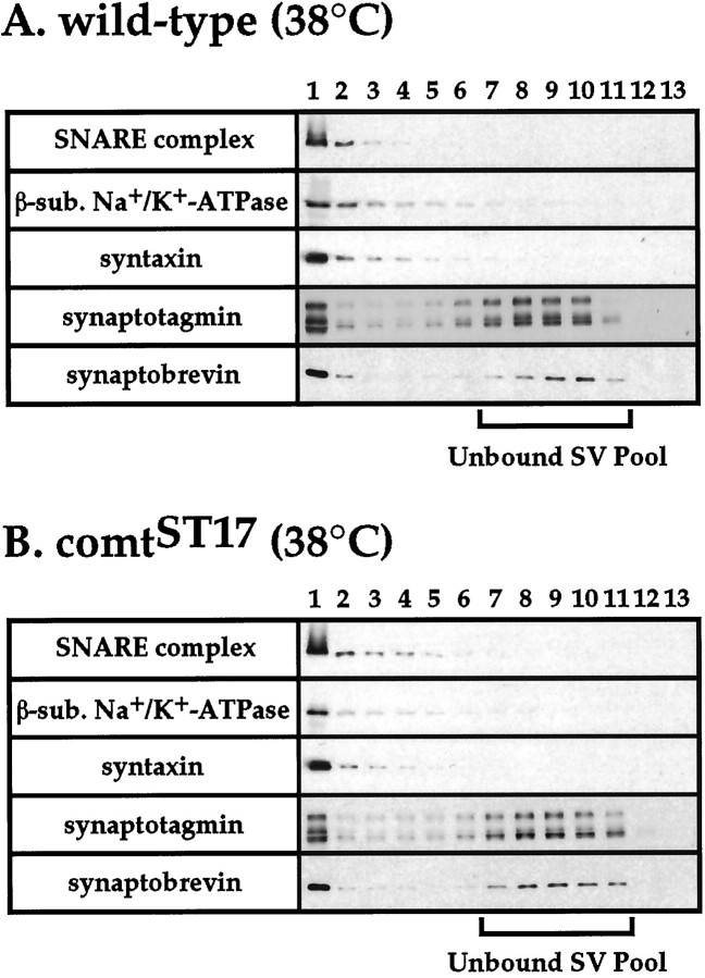
Fig. 4.
The SNARE complex resides in the plasma membrane. A postnuclear head homogenate from wild-type flies (A) or comtST17mutants (B) was sedimented on a glycerol gradient, and aliquots of the gradient fractions (numbered 1–13 from the bottom to the top of the gradient) were subjected to Western blot analysis to identify peak plasma membrane fractions, peak synaptic vesicle fractions, and SNARE complexes. Western blot analyses were performed using antisera to a 42 kDa plasma membrane marker [the putative β-subunit of the Na+/K+ ATPase, which is recognized by rabbit antisera to horseradish peroxidase (van de Goor et al., 1995)] and to the synaptic vesicle proteins n-synaptobrevin (a mixture of the NSYB1 and NSYB2 antisera), synaptotagmin (DSYT2) (Littleton et al., 1993), and synapsin (data not shown), as well as a monoclonal anti-syntaxin antibody to detect SNARE complexes and monomer syntaxin. Analyses to identify syntaxin- and n-synaptobrevin-containing gradient fractions were performed using samples boiled 5 min to disrupt SNARE complexes. SNARE complex-containing gradient fractions were detected using unboiled samples. No comparison of SNARE complex abundance can be made between A and Bbecause immunoblot exposure times varied in these experiments. Results obtained with the anti-synapsin antiserum were consistent with those obtained with antisera to the other synaptic vesicle antigens analyzed. Results using flies exposed to restrictive temperature (38°C) for 20 min before preparation of head homogenates are shown. Similar results were obtained using flies maintained at permissive temperature (22°C).

