
Fig. 5.
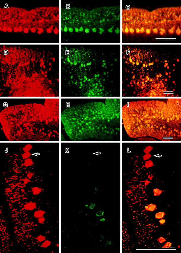
Dual immunofluorescence staining of parasagittal sections from mutant cerebella at P24 by anti-calbindin (red) and anti-c-fms(green) antibodies. The regions expressing both c-fms and calbindin are visualized asyellow regions. In osteopetrotic (A–C), reeler (D–F), and weaver (G–I) mutant cerebella, the patterns of expression of c-fms and calbindin are similar. Displaced Purkinje cells in the reeler mutant express c-fms. Some of the staggerer Purkinje cells (J–L) do not express c-fms (arrows). Scale bars, 100 μm.