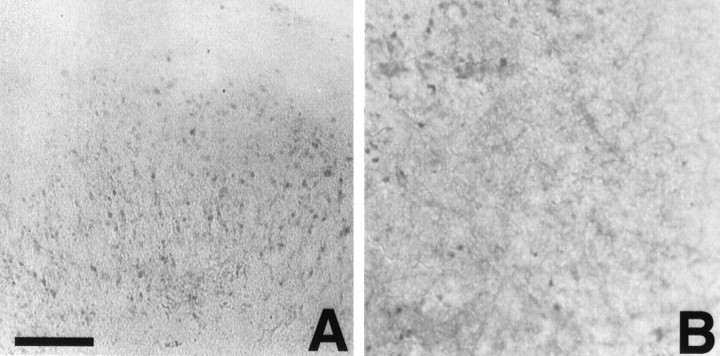
Fig. 5.
Aβ staining that uses end-specific antibodies for Aβ40 and Aβ42. A, Aβ42 staining in the CA1 pyramidal region of the slice culture after treatment with Aβ plus TGF-β2. B, Aβ40 staining in the stratum radiatum region of a slice treated with Aβ plus TGF-β1. Most of the Aβ40 is diffuse with some apparent microglial staining, whereas the Aβ42 staining is predominately cellular. Scale bar, 40 μm.

