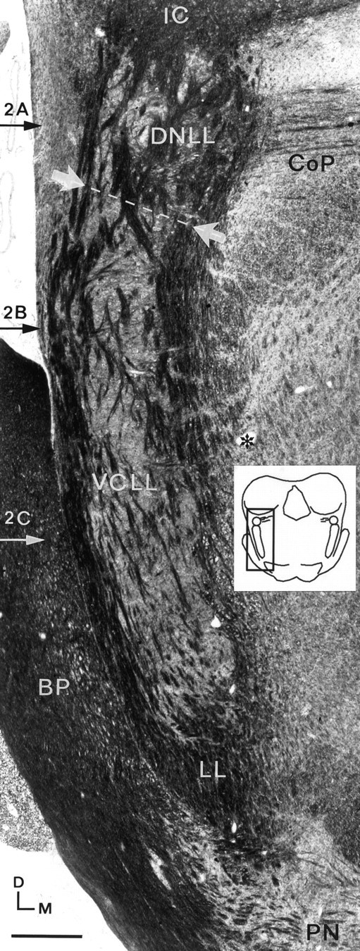
Fig. 1.

Photomicrograph of a Woelcke- and cresyl violet-stained frontal section through the cat brainstem.Horizontal arrows labeled 2A–2C indicate the approximate levels of the corresponding horizontal sections shown in Figure 2. The border between the DNLL and VCLL is indicated withwhite arrows and a dotted line. The lemniscal fibersencapsulate and pierce the VCLL and the DNLL in a continuous system. The lemniscus is supplied medially by ascending auditory fibers from the superior olivary complex and the dorsal cochlear nucleus crossing the midline in the reticular formation (asterisk). The thinner fiber fascicles of the commissure of Probst pierce the lemniscus from the medial side at the level of the DNLL. Scale bar, 500 μm. BP, Brachium pontis; CoP, commisure of Probst; D, dorsal; LL, lateral lemniscus; M, medial;PN, pontine nuclei.
