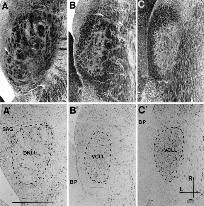
Fig. 2.

Photomicrographs of pairs of adjacent horizontal sections through the lateral lemniscus at the correspondingly labeled levels in Figure 1. The sections in each pair have been stained for myelin and cells (Woelcke and cresyl violet; A–C) and cells only (thionin; A′–C′). A andA′ are at the level of the DNLL; the others are through the VCLL. The cellular area, inside the tube of external fibers, is marked by a dotted line in A′–C′. Differences in cytoarchitecture between the DNLL and VCLL have been used as criteria for defining the border between the two in the present study. The DNLL (A′) is seen to be populated by uniformly large cells, which occur in irregular groups between the thick, unstained fiber fascicles. The VCLL (B′, C′), in contrast, appears to contain neurons of various sizes, mainly medium-sized. The cells are more evenly dispersed, particularly in the ventral part where the cell density is also larger (C′), in conformity with the less prominent internal fiber component (C). Scale bar, 1 mm. BP, Brachium pontis; L, lateral; R, rostral;SAG, sagulum.
