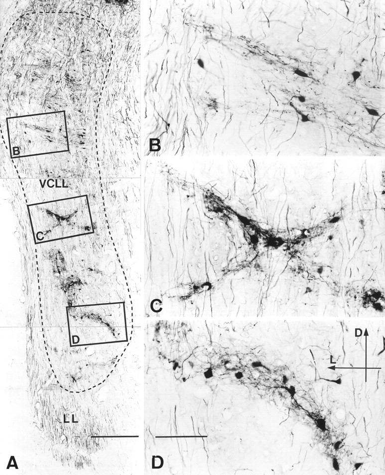
Fig. 5.

Photomicrographs of a transverse section through the VCLL of cat 94070 illustrating the labeling after BDA injection into the middle-frequency region of the ipsilateral IC (compare Fig.3B). The framed regions are shown at higher magnification in B–D. The labeled structures are distributed in 2-D patches (3-D clusters) composed of labeled cell bodies, proximal dendrites, and a terminal fiber plexus. The patches are shaped as irregular bands ∼150 μm thick. Elongated, labeled cell bodies and primary dendrites appear oriented in parallel with the long axis of the bands. Labeled axons en passage course vertically throughout the tissue. Dorsally, the labeling is more diffuse. Scale bars: A, 1 mm; B–D, 200 μm.D, Dorsal; L, lateral. LL, lateral lemniscus.
