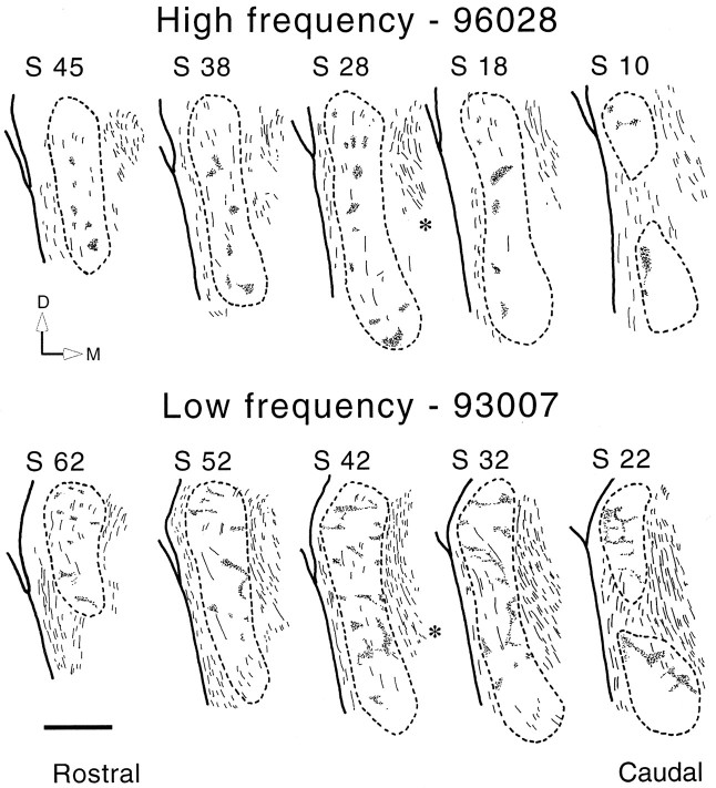
Fig. 6.
Series of camera lucida drawings illustrating the patches of labeling in the VCLL after injections in the high-frequency region (compare Fig. 3A) and the low-frequency region (compare Fig. 3C) of the ipsilateral CNIC. Asterisks indicate lemniscal fibers crossing in the reticular formation (compare Fig. 1). In the high-frequency case, most of the labeling is located in the lateral half of the VCLL. The low-frequency case shows a larger amount of labeling. In this case, the patches occur throughout most of the VCLL, and topographic differences are difficult to define. The central part of the VCLL, however, contains less labeling than the medial and lateral borders. Scale bar, 1 mm. D, Dorsal;M, medial; S, section number.

