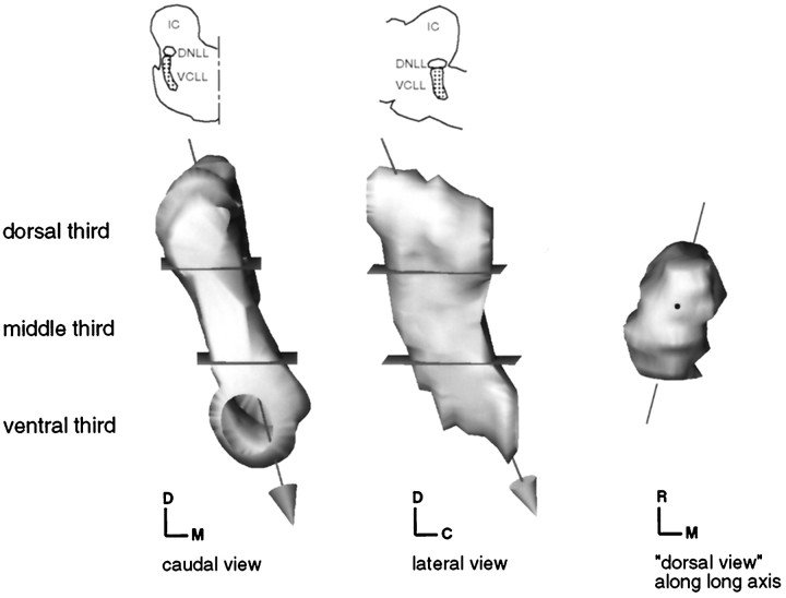
Fig. 7.
Computer-generated surface model of the VCLL, showing how the VCLL arbitrarily has been divided into dorsal, middle, and ventral thirds. The arrow represents the long axis of the VCLL. The angle of views from caudal, lateral, and dorsal (along the long axis of the VCLL), correspond the views used in Figures 8-12.C, Caudal; D, dorsal; M, medial; R, rostral.

