Fig. 6.
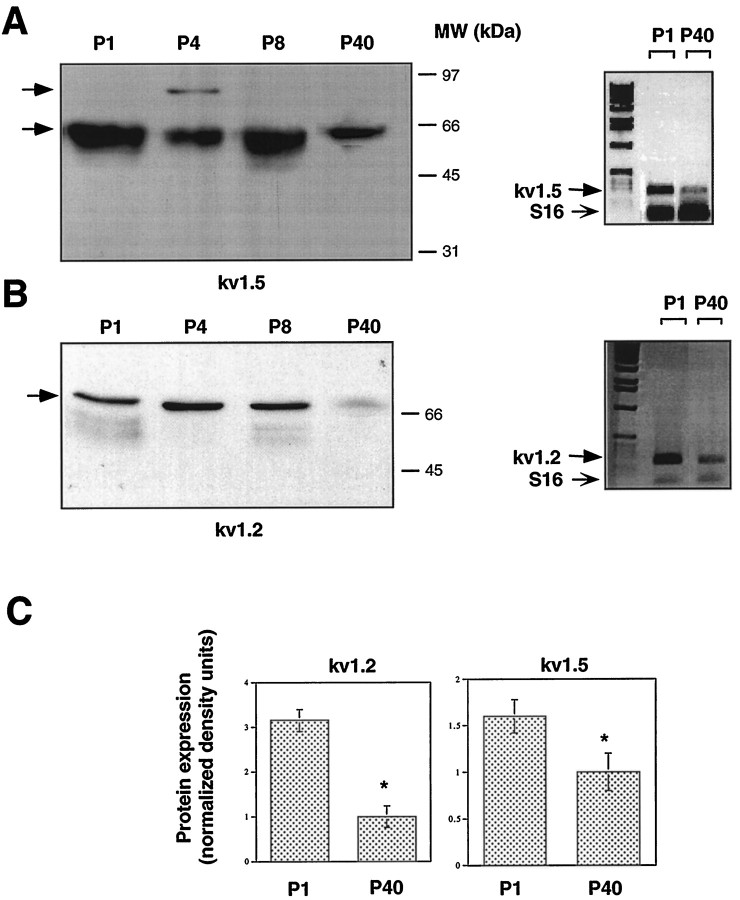
Expression of Kv1.2 and Kv1.5 channel α subunits in mouse sciatic nerve during postnatal development. A,Left, Membrane fractions of sciatic nerves from P1, P4, P8, and P40 mice were subjected to SDS-PAGE and immunoblot analysis with anti-Kv1.5 antibodies. To estimate and compare total protein inputs in each lane, blots were stained with Ponceau S before immunoblot analysis (data not shown). Right, RT-PCR, followed by Southern blot analysis of Kv1.5 transcripts in sciatic nerves from P1 and P40 mice. B, Left, Immunoblot analysis of Kv1.2 on postnatal sciatic nerve as inA. Right, RT-PCR and Southern blot analysis of Kv1.2 as in A. Primer pairs to the specific 3′ coding regions of either Kv1.5 (A) or Kv1.2 (B) amplified PCR fragments of 273 and 248 bp, respectively. The bottom band represents the S16 ribosomal protein PCR fragment (102 bp), which was used to estimate the starting input RNA. C, Quantitation of the developmental downregulation of Kv1.2 and Kv1.5 proteins from P1 to P40 sciatic nerve as illustrated in A and B. Data of densitometric scanning were normalized to values of P40 and represent mean ± SEM of three independent experiments. *p < 0.05, differs significantly from P40.

