
Fig. 2.
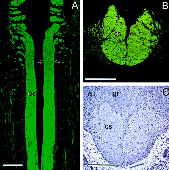
The normal rat corticospinal tract (cs) in horizontal section (A) and cross section (B, C, at the level of the lesions and transplants; arrows in A). A, B, CaMII; C, NF. cu, Cuneate fasciculi; gr, gracile fasciculi; x, fibers leaving the pyramidal decussation. Scale bars, 500 μm.