
Fig. 10.
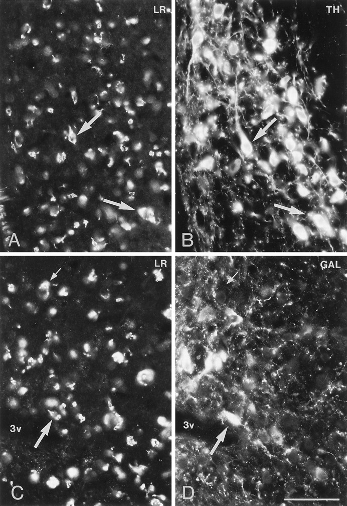
Immunofluorescence photomicrographs of sections of the arcuate nucleus (Arc) after direct double labeling combining antiserum to LR (A, C) with antiserum to tyrosine hydroxylase (TH) (B) or galanin (GAL) (D). Scale bars, 50 μm.
Official websites use .gov
A
.gov website belongs to an official
government organization in the United States.
Secure .gov websites use HTTPS
A lock (
) or https:// means you've safely
connected to the .gov website. Share sensitive
information only on official, secure websites.
Immunofluorescence photomicrographs of sections of the arcuate nucleus (Arc) after direct double labeling combining antiserum to LR (A, C) with antiserum to tyrosine hydroxylase (TH) (B) or galanin (GAL) (D). Scale bars, 50 μm.