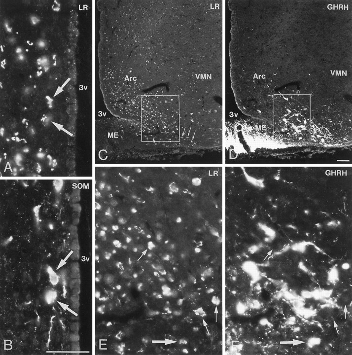
Fig. 11.

Immunofluorescence photomicrographs of sections of the periventricular nucleus (A, B) and arcuate nucleus (Arc) (C–F) after direct double labeling combining antiserum to LR (A, C, E) with antiserum to somatostatin (SOM) (B) or growth hormone-releasing hormone (GHRH) (D, F). Some LR-IR cells of the periventricular nucleus are SOM-positive (arrows). LR-IR neurons are distributed mainly in the ventromedial part of the Arc, whereas GHRH-containing neurons are mainly present in the ventrolateral part (C, D). Some neurons in the far ventrolateral part contain both LR- and GHRH-LI (small arrows in C, D). There are many LR-IR neurons that are GHRH-negative (small arrows inE, F) and many GHRH-positive neurons that are LR-negative. Few neurons contain both LR- and GHRH-LI (large arrow in E, F). ME, Medan eminence; 3v, third ventricle. Scale bars, 50 μm.
