
Fig. 2.
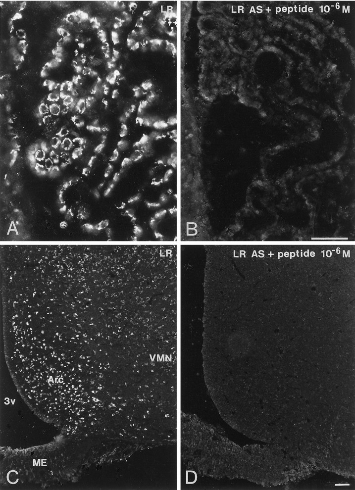
Immunofluorescence photomicrographs of sections of the choroid plexus (A, B) and hypothalamic arcuate nucleus (C, D) after incubation with antiserum to LR (A, C) or antiserum to LR preadsorbed with immunogen peptide (10−6m) (B, D). Strong LR-LI is present in the periphery of cells in the choroid plexus (A). In the arcuate nucleus (Arc), strong LR-LI is seen mainly in the ventromedial part of the nucleus but also in some neurons belonging to the dorsomedial and ventromedial part. Weaker LR-LI is present in the ventromedial nucleus (VMN). Note total absence of LR-LI in sections incubated with preadsorbed antiserum.ME, Median eminence; 3v, third ventricle. Scale bars, 50 μm.