
Fig. 3.
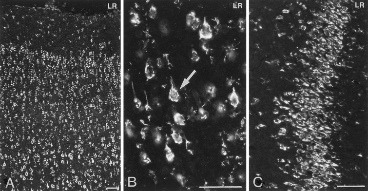
Immunofluorescence photomicrographs of sections of the rat cerebral cortex (A, B) and the CA3 region of the hippocampus (C) after incubation with antiserum to LR. Strong LR-LI is present in neurons in layers II–VI of the cerebral cortex (A) and in neurons of the CA3 region of the hippocampus. At higher magnification (B), it can be seen that LR-LI is present in the periphery of pyramidal neurons (see arrow inB). Scale bars, 50 μm.