
Fig. 6.
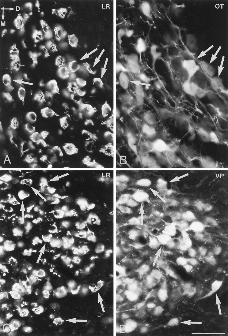
Immunofluorescence photomicrographs of sections of the supraoptic nucleus (A, B) and the magnocellular part of the paraventricular nucleus (C, D) after direct double labeling combining antiserum to LR (A, C) with antiserum to oxytocin (OT) and vasopressin (VP). Comparison of A withB and C with D shows that LR-LI is present in both VP- and OT-containing neurons (arrows). D, Dorsal direction;M, medial direction. Scale bar, 50 μm.