
Fig. 9.
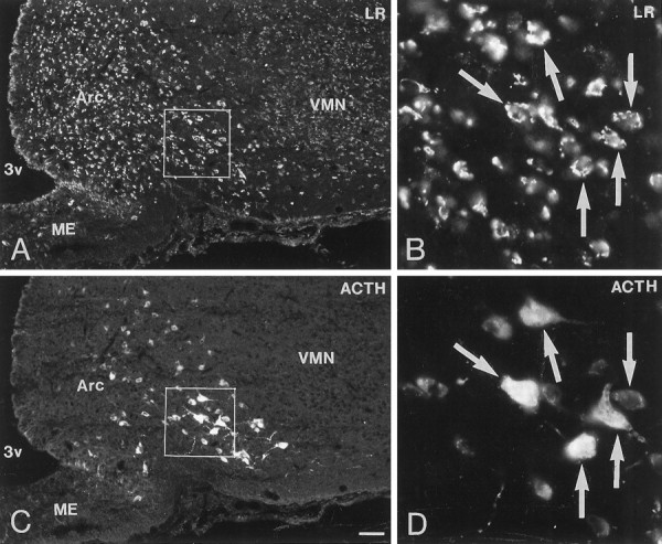
Immunofluorescence photomicrographs of sections of the arcuate nucleus (Arc) after direct double labeling combining antiserum to LR (A, C) with antiserum to adrenocorticotropic hormone (ACTH) (B, D). Large ACTH-positive POMC-containing neurons are distributed in the ventrolateral part of the Arc. Double labeling shows co-localization of LR- and ACTH-LI in many neurons (arrows). Scale bar, 50 μm.