
Fig. 2.
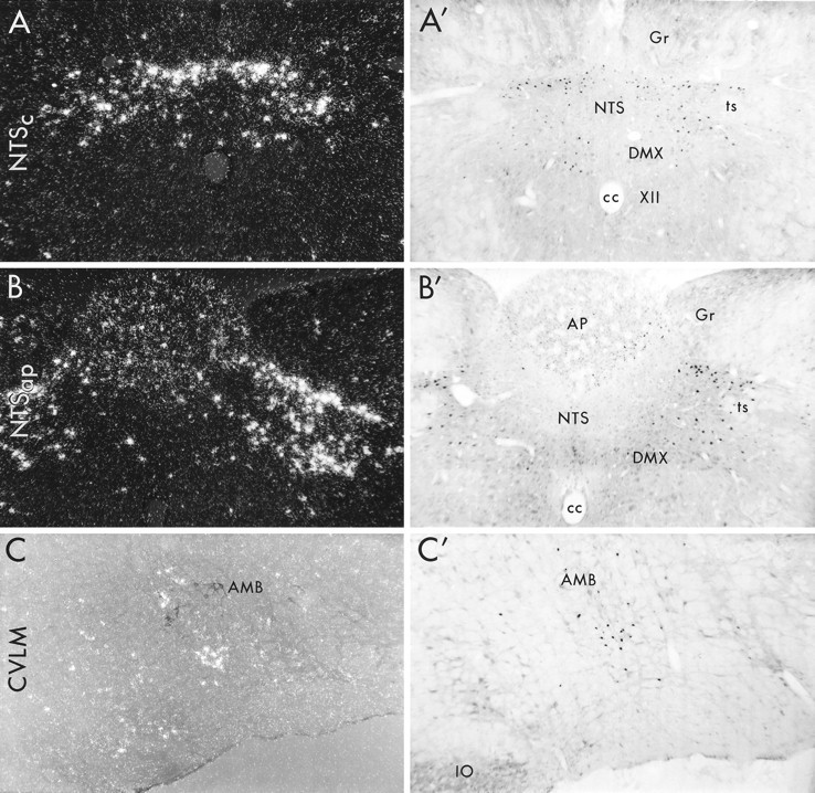
Medullary patterns of c-fos mRNA and Fos protein expression induced by prolonged PE infusion. Dark field (A, B) and bright field (A′, B′, C′) photomicrographs depicting hybridization and avidin–biotin immunoperoxidase localization of c-fos mRNA and Fos-IR in the NTS at the levels of the commissural subnucleus (NTSc; A,A′) and the area postrema (NTSap; B,B′) and in the CVLM (C,C′) at 1 and 2 hr after hypertensive challenge, respectively. C, Combined immuno- and hybridization histochemical detection of choline acetyltransferase-IR cells in the nucleus ambiguus (black cells) and PE-inducedc-fos mRNA (white silver grains), respectively, photographed using polarized epifluorescence illumination. In contrast to the acute infusion model, sustained hypertension resulted in comparable strengths and distributions ofc-fos mRNA and protein expression in each region. This includes robust induction in the dorsal baroreceptor strip of the commissural NTS (NTSc) that extended into the dorsal and dorsolateral aspects of NTS at the level of area postrema (NTSap). Noncatecholaminergic neurons of the CVLM region were also reliably activated.AMB, Nucleus ambiguus; AP, area postrema;cc, central canal; DMX, dorsal motor nucleus of vagus; Gr, gracile nucleus;IO, inferior olive; NTS, nucleus of solitary tract; ts, solitary tract; XII, hypoglossal nucleus. All photomicrographs, 75×.