Fig. 5.

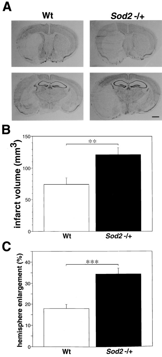
Histological analysis after 24 hr of MCA occlusion. A, Photomicrograph showing the histological changes after 24 hr of MCA occlusion in wild-type (Wt) and knock-out mutant mice (Sod2 −/+). The infarct area was localized in the caudoputamen and MCA territory cortex in both mice groups. However, cortical infarction extended to the boundary zone of the anterior cerebral artery territory, and brain swelling was extremely severe in the knock-out mutant mice. Also shown are infarct volume (B) and hemisphere enlargement (C) in wild-type and knock-out mutant mice at 24 hr ischemia. Values are mean ± SE; **p < 0.01 and ***p < 0.001, Student’s ttest. Cerebral infarction and hemisphere enlargement were significantly more severe in knock-out than in wild-type mice. Scale bar, 1 mm.
