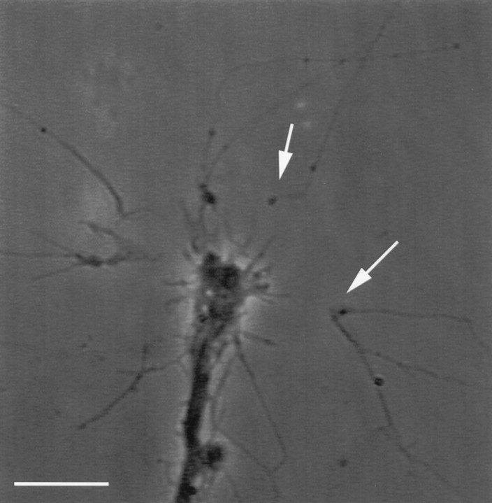
Fig. 4.
Surgical isolation of individual filopodia. Isolated filopodia (arrows), often 20–30 μm long, exhibit intact morphology after laser-assisted transection from their parent, fura-loaded growth cones. A large gap forms between an isolated filopodium and parent growth cone after laser-assisted transection (arrows). This gap results from a retraction of each of the cut ends. This phase picture of a fura-loaded growth cone (5 μm fura-2AM) was taken 20 min after the surgical isolation of filopodia. Scale bar, 10 μm.

