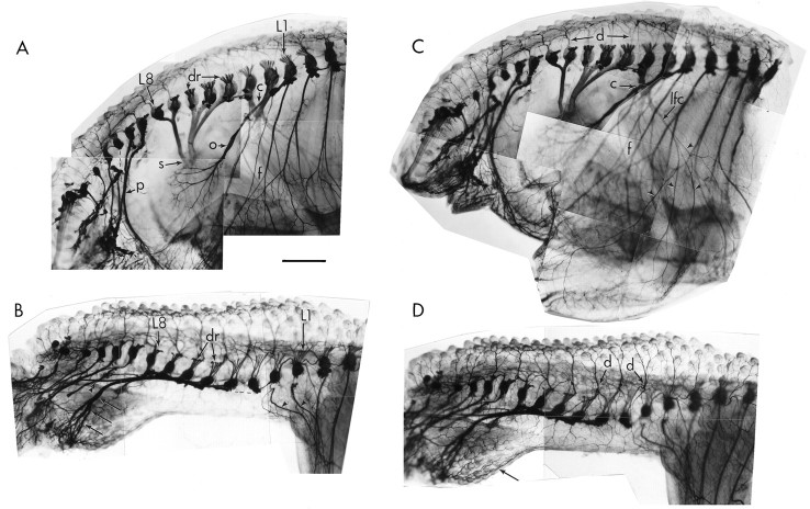
Fig. 8.
Peripheral nerve patterns in whole mounts of normal and experimental limbs as revealed by staining with the TUJ1 antibody. A and B are focused on the medial aspect of each preparation to show nerve projection patterns;C and D are focused on the lateral aspect to show skin innervation in the same limbs. A, Normal pattern of peripheral nerve projections. Lumbar segments L1–L3 largely contribute axons to the crural plexus (c), which gives rise to many peripheral nerves of the limb including the prominent obturator nerve (o). Lumbar segments L3–L8 all contribute axons to the sciatic plexus (s). The more caudal segments contribute axons to the pudendal plexus (p). f, Femur.B, After limb-bud deletion, neither the crural plexus nor the sciatic plexus develops. L1 and L2 typically project axons anteriorly toward thoracic segments (arrowheads). L2–L8 all project axons posteriorly toward the tail, where they form a novel plexus (arrows) and contribute to the pudendal plexus (double arrowhead). Dashed lines indicate connections broken during processing; dr indicates dorsal roots. The spinal cord and sympathetic chains were removed for clarity. C, Normal patterns of skin innervation on the lateral surface of the thigh. The crural plexus (C) gives rise to the lateral femoral cutaneous nerve (lfc), which branches (arrowheads) to provide much of the cutaneous innervation of the lateral thigh. The more proximal skin receives axons from the dorsal rami (d) of each spinal nerve. f, Femur. D, After limb-bud deletion, the lateral femoral cutaneous nerve does not develop, and the remaining skin is innervated by branches from dorsal rami (d). Some axons also reach the skin over the deletion site from the novel plexus in the tail region (arrow). Scale bar, 1 mm.

