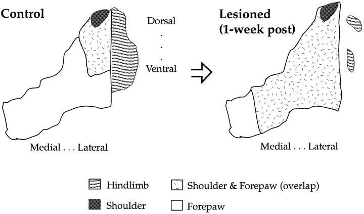
Fig. 11.
Schematic of the topographic overlap between shoulder and forepaw in the focal zone at 1 week and 1 month after lesion. This is a coronal view of forepaw, shoulder, and hindlimb sensory maps in the VPL. The plasticity depicted is derived from data on the volume of shoulder–forepaw overlap (Fig. 2) and planar analysis (Figs. 7, 8).

