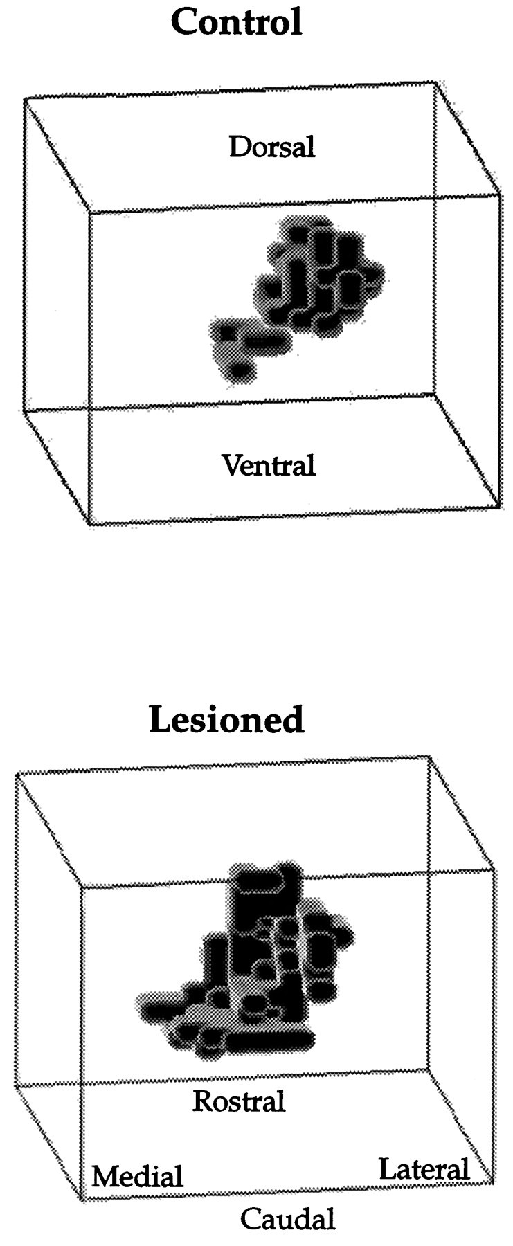
Fig. 4.

Spatial distribution of recording sites responsive to shoulder stimulation from control and 1 week postlesion animals. This is a caudal perspective of the sensory map. Lesion-induced changes are most marked at the rostral pole, toward the anterior end of the display. Much of the shoulder expansion appears to occur along the mediolateral axis after gracilis lesions.
