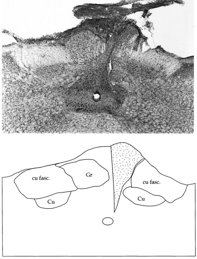
Fig. 9.

Photomicrograph depicting a coronal view of the dorsal column nuclei stained with Nissl 1 week after lesion. The damage observed in the gracile nucleus existed along the length of the nucleus. The accompanying diagram is a schematic rendering of the photomicrograph, barring tissue-processing artifacts. The zone of gliosis is indicated by the stippled area. cu fasc, Cuneatus fasciculus; Cu, cuneate nucleus;Gr, gracile nucleus.
