
Fig. 1.
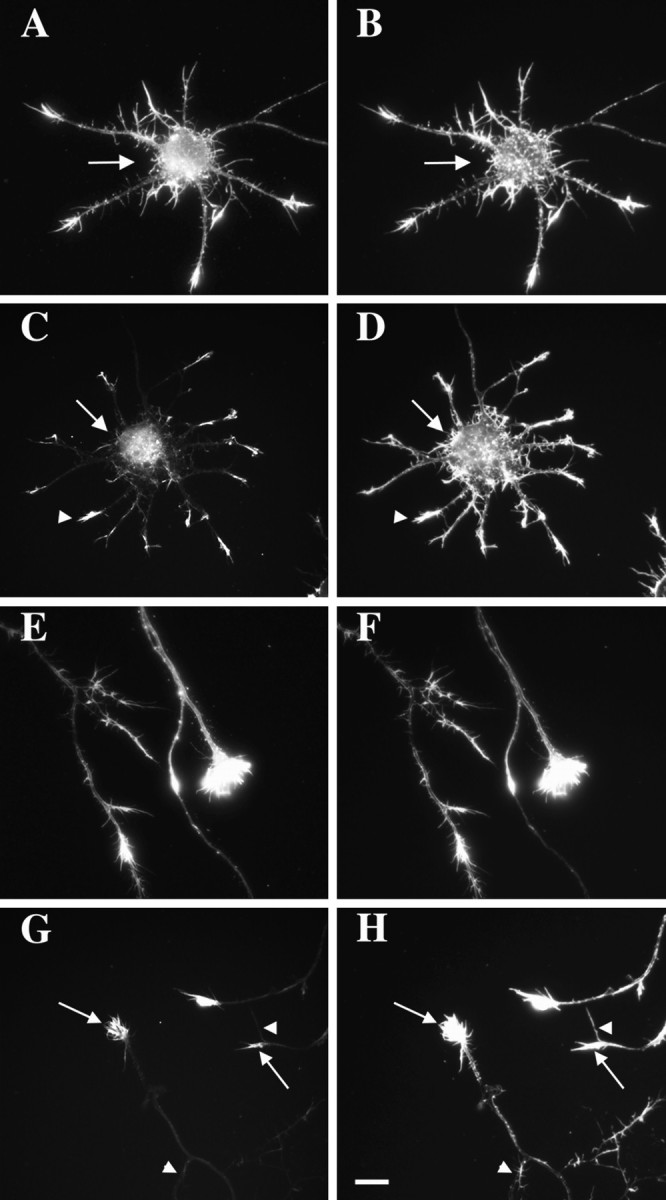
Localization of actin isoforms within cultured neurons. Cortical neurons cultured for 4 d were double-labeled with an isoform-specific antibody to β-actin or γ-actin (left column) and phalloidin–TRITC (right column). A, B, γ-Actin was distributed throughout the cell body (arrow) and neurites and resembled phalloidin staining. C,D, Localization of β-actin at tips of minor neurites (arrowhead). Low levels of β-actin were observed in the cell body (arrow). Phalloidin labeled actin filaments throughout the cell body (arrow) and minor neurites (arrowhead). E,F, γ-Actin was distributed throughout the axon and growth cone, as was phalloidin staining. G,H, β-Actin was enriched within axonal growth cones (arrows). Only weak labeling was observed in the axon shaft. Not all filopodia were labeled (arrowheads) despite the presence of F-actin (phalloidin). Scale bar, 8.5 μm.