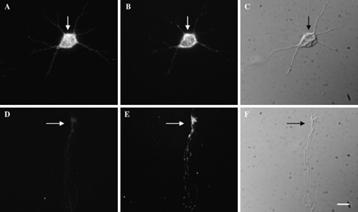
Fig. 4.
Intraneuronal distribution of β-actin and γ-actin mRNA. Cortical neurons were cultured for 4 d, at which time most neurons have distinguishable axonal and dendritic processes.A, Hybridization of biotinated probes to γ-actin mRNA within the cell body (arrow). B, Hybridization of digoxigenin-labeled probes to β-actin mRNA within the cell body (arrow). C, Differential interference contrast (DIC) microscopy of the cell body (arrow), minor neurites, and a single axon. The axon is considerably longer than the minor neurites and cannot be photographed in entirety at this magnification. Shown here is the initial segment.D, Absence of γ-actin mRNAs from the axonal growth cone (arrow). E, Localization of β-actin mRNA within the axonal growth cone (arrow).F, DIC image of axonal growth cone (arrow) from this axon. The axon in this cell is ∼150 μm in length. Scale bar, 10 μm.

