
Fig. 6.
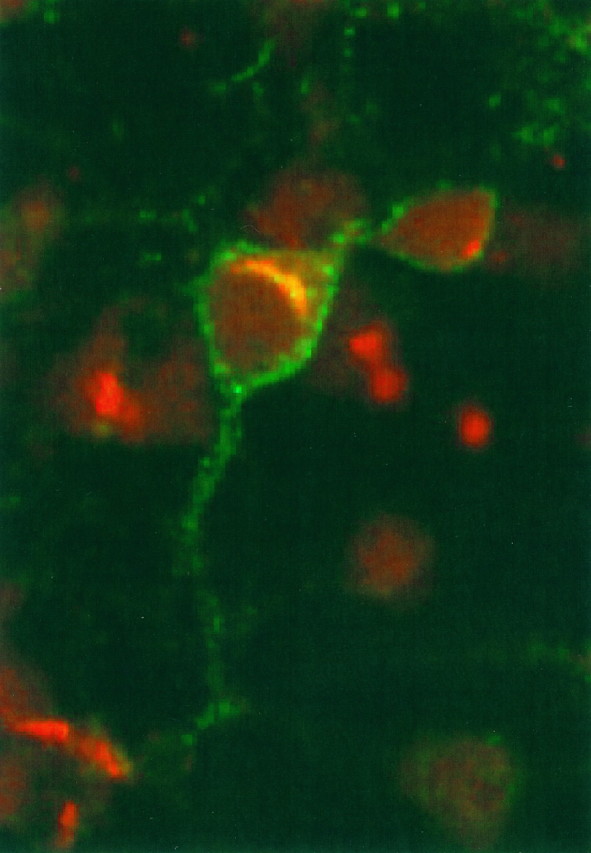
Double label, confocal microscopy demonstrates that intracellular Cat-315 immunoreactivity localizes to the Golgi complex. E16 plus 3 CD cultures were fixed, permeabilized, and then double labeled with Cat-315 (green) and a polyclonal antibody that recognizes the Golgi apparatus (red). Cat-315 immunoreactivity is distributed along the neuronal cell surface (green surface staining). The intracellular Cat-315 signal appears yellow, because of signals from both antibodies. In every case, intracellular Cat-315 immunoreactivity precisely colocalizes with immunoreactivity for the anti-Golgi antibody (yellow). However, there are many cells with Golgi labeling but without Cat-315 labeling (red). The figure shows a 2-μm-thick confocal image, in which antibodies were visualized with FITC- or Texas Red-conjugated, species-specific secondary antibodies.