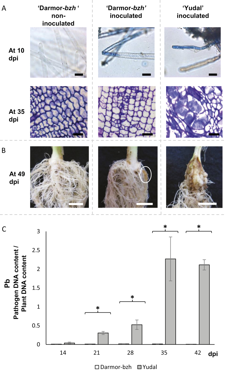
Fig. 1.
Time course of compatible interactions in the two genotypes of B. napus, Darmor-bzh and Yudal, inoculated with the eH isolate of P. brassicae. (A) Histopathological characterization of clubroot infection in the root hairs at 10 days post-infection (dpi) and from transverse sections of the roots at 35 dpi of inoculated and non-inoculated plants. Staining with 1% toluidine blue allows visualization of P. brassicae inside root hairs during the primary phase, and inside cortical cells during the secondary phase. Bars=25 μm. (B) Typical root symptoms at 49 dpi. Bars=1 cm. The white ellipse on the central picture indicates the presence of a small gall. (C) Dynamics of pathogen root invasion in Darmor-bzh and Yudal followed by quantitative PCR using DNA extracted from whole infected roots. The internal transcribed spacer region of P. brassicae (PbITS) was amplified and compared with the Cruciferin A gene of B. napus (BnCruA), and the ratio of pathogen to plant DNA (Pb) was calculated. Samples were analysed at 14, 21, 28, 35, and 42 dpi. Data are expressed as the mean ±SE of four replicates. Asterisks indicate significant differences in Pb values between Darmor-bzh and Yudal at the same sampling time (P<0.05, Wilcoxon-Mann-Whitney test). (This figure is available in colour at JXB online.)

