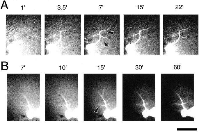
Fig. 4.
Penetration of IgG into Purkinje cells from patch pipette. Purkinje cells were labeled with FITC-labeled goat IgG. Theabscissa indicates the time after break-in. The IgG reached the secondary and tertiary dendritic regions (arrowheads) of the Purkinje cell within 3.5 min after patch formation (A), and the fluorescence intensity did not increase much more after the 15 min time point. Each set of images was taken and displayed with the same exposure and display conditions in A and B. A confocal laser scanning unit was used in A but was not used inB. Scale bar, 50 μm.

