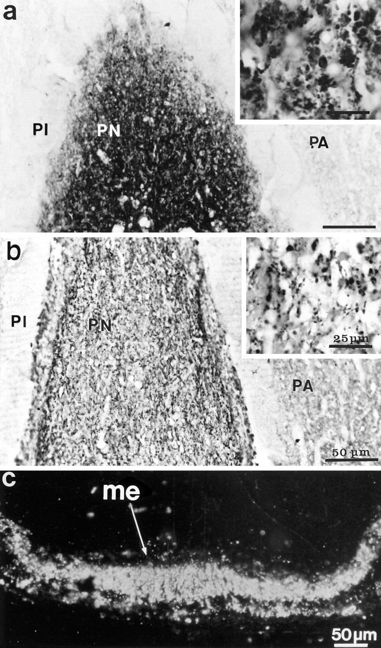
Fig. 3.

F3 immunoreactivity in the hypophysis of normal (a) and dehydrated (b) adult rats. Labeling is restricted to the posterior lobe (PN), or neurohypophysis, where it appears associated with fibers and dilatations (shown at higher magnification in the insets). The reaction is greatly diminished in glands of stimulated rats (b). Immunoperoxidase labeling was revealed with DAB and viewed with bright-field optics.PA, Pars anterior; PI, pars intermedia.c, F3 immunoreactivity in the median eminence (me) of a dehydrated rat. Immunoperoxidase labeling (here illustrated with dark-field optics) revealed a strong signal, regardless of the condition of the animal, throughout its internal layer, where the HNS tract courses.
