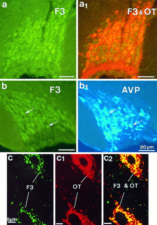
Fig. 5.

Simultaneous immunofluorescent localization of F3 and oxytocin (OT) or vasopressin (AVP) immunoreactivities in the SON. F3 was visualized with FITC-conjugated (green) antibodies; OT and AVP were visualized with Texas Red and AMCA-conjugated (blue) antibodies, respectively. a, b, Light microscopy of frozen frontal sections (25 μm) showed F3 immunoreactivity (green, a, b) in magnocellular somata and fibers throughout the nucleus, colocalized with OT (orange, a1) or AVP (arrows,b and b1). Epifluorescence with appropriate filters was used. c, c2, Confocal microscopy of a semithin (700 nm) frozen section of the SON simultaneously immunolabeled for F3 (green, c) and OT (red, c1). In c and c1, each image represents a single optical projection. Note that the labeling for F3, as for OT, is punctate and is dispersed throughout the cytoplasm of somatic and dendritic profiles. In c2, thered–green overlay of the two antigens clearly shows colocalization (yellow) of the two immunoreactivities over the same punctae. Note that immunolabeling caused by OT is more extensive than that caused by F3. The better resolution afforded by these sections shows clearly that labeling for F3, as for the neuropeptide, is intracytoplasmic; no reaction is visible on either somatic or dendritic surfaces.
