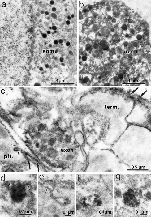
Fig. 6.

Electron micrographs depicting F3 immunoreactivity in noncounterstained ultrathin sections of the SON (a, b) and neurohypophysis (c–g) of adult rats after preembedding immunoperoxidase staining. Electron-dense peroxidase reaction product representing F3 immunoreactivity covers secretory granules in the cytoplasm of somatic (a) and axonal (b, c) profiles. Labeling of neuronal surfaces is absent in the SON (a, b) but is present on the surfaces of axon terminals (term.) and glial cells in the neurohypophysis (c–e). Note that the reaction product is associated with invaginations (arrows) of glial (c) and terminal (d, e) surfaces and with multivesicular bodies (f, g) in axon terminals. In the neurohypophysis (c), reaction also occurs in extracellular spaces (large arrows).
