
Fig. 2.
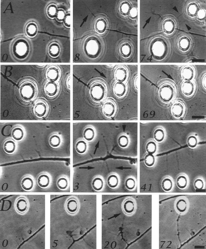
Axonal motile responses at sites of NGF bead contact. Although filopodial sprouts that are formed on the bead surface are not visible using phase-contrast optics, some filopodia extended beyond beads. The numbers in the panels refer to minutes after the first image in the series. A shows a filopodium that extended from the site of axon–bead contact (arrow, 0–8 min) and developed into a collateral branch with a small growth cone-like structure at its tip (arrow, 8–74 min). Two other filopodia also extended from the axon and contacted a nearby bead (arrowheads, 74 min). Filopodia, generated underneath beads, that contacted additional beads became stabilized (B,arrow at 5–69 min). C shows a region of an axon ∼50 μm behind the growth cone (data not shown) that became spontaneously active, generating filopodia that contacted beads and were stabilized (arrows, 3–41 min). One filopodium thickened as the axon translocated toward the bead it contacted (arrowhead, 3 min), because of the activity of the leading growth cone, and eventually came to rest underneath the bead. Spontaneously formed collateral branches that contacted NGF-coated beads turned and extended toward the beads after initial contact (D). Scale bar, 10 μm.