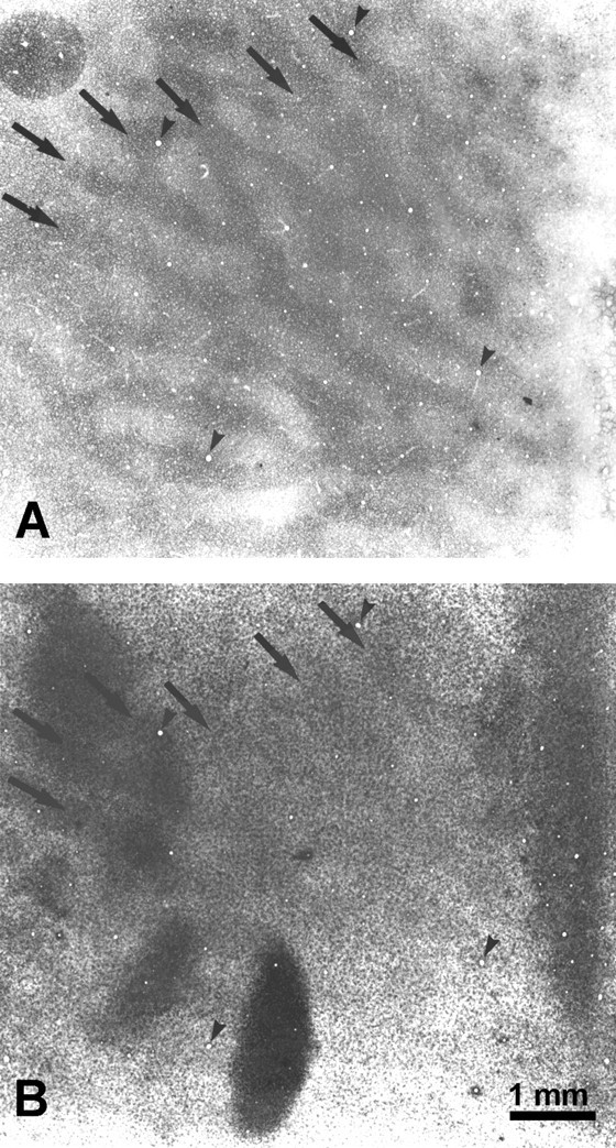
Fig. 10.

Monkey 7 (laser vs normal). Boxed region from Figure 9, comparing the CO (A) and Zif268 (B) patterns inside the cortical scotoma. The dark columns match (arrows), indicating that higher Zif268 levels persisted in the right eye’s ocular dominance columns, despite 10 hr of dark adaptation, enucleation of the right eye, and 4 hr of stimulation of the left eye. Presumably the laser lesion prevented visual stimulation of the left eye from inducing greater Zif268 levels in its ocular dominance columns. The section contains some blotchy artifact, perhaps from tissue manipulation during flat mounting.
