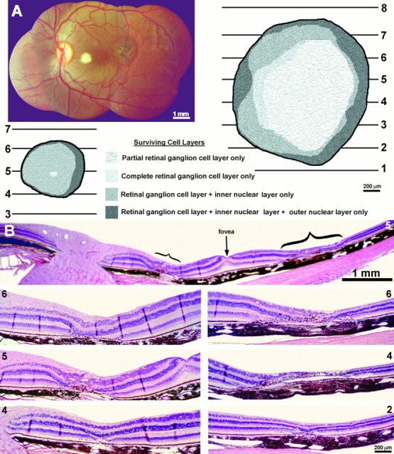
Fig. 11.

Monkey 8 (laser vs enucleation).A, Montage of the left retina, prepared from photos taken immediately after applying 15 laser spots at a setting of 180 mW to create a 0.5 mm lesion 6–9° nasal to the fovea. The 1.5 mm lesion, 6–12° temporal to the fovea, was made 14 weeks earlier by applying 25 spots at 220 mW. Note the dramatic difference in the appearance of the fresh lesion and the old lesion. Thediagrams show the extent of retinal damage from the two lesions, compiled from serial paraffin sections. B, Sample histological sections show the damage from the smaller nasal (bottom left) and larger temporal (bottom right) laser lesions. The temporal lesion caused more severe inner retinal damage and had more sloping borders. Both these features were reflected in the cortical findings illustrated in Figure 12.
