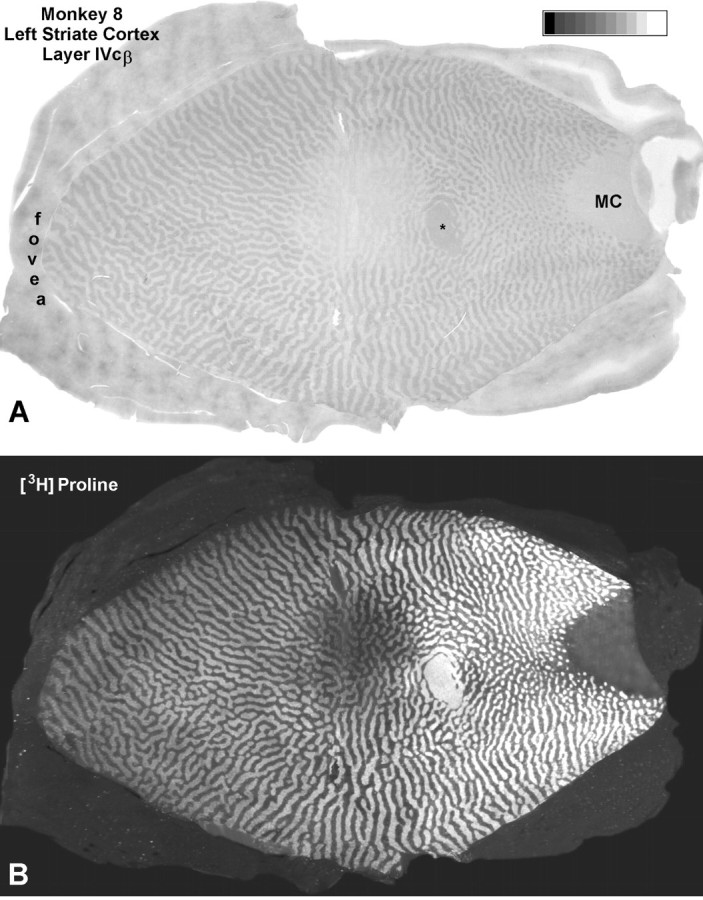
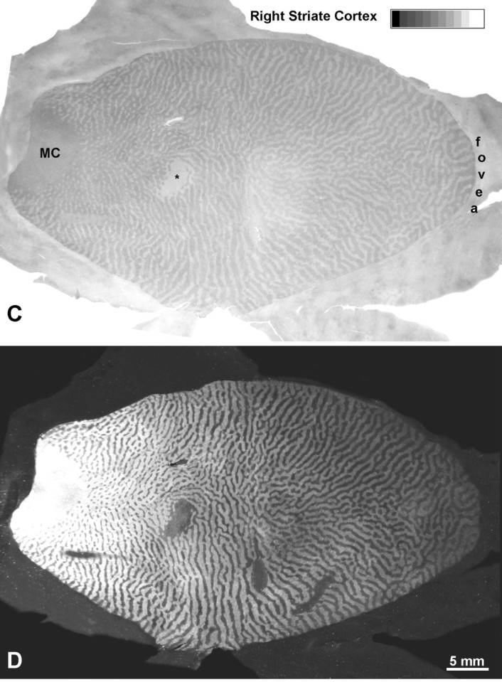
Fig. 12.


Monkey 8 (laser vs enucleation).A, CO montage showing the cortical scotoma induced by the laser lesion temporal to the fovea in Figure 11, silhouetted against a backdrop of ocular dominance columns induced by enucleation of the right eye. Note the gradual loss of CO activity from the periphery to the center of the scotoma. Even in the center, CO activity is slightly greater in the ocular dominance columns of the lesioned eye than in those of the enucleated eye. MC, Monocular crescent; *blind spot. B, Hole in the autoradiographic montage caused by laser injury to ganglion cells. It also has sloping edges but appears shallower than the metabolic hole inA, because columns are still seen clearly throughout.C, Montage of the scotoma resulting from the laser lesion nasal to the fovea. Because the laser damage was less severe and more evenly distributed, the loss of CO activity is milder and lacks a periphery-to-center gradient. D, Sparing of ganglion cells was confirmed by 3[H]proline labeling within the CO scotoma, which remained nearly normal. This experiment shows that outer retinal damage reduces cortical CO activity, but less than inner retinal damage or enucleation.
