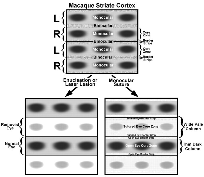
Fig. 17.
Schematic diagram of normal macaque striate cortex (top), looking through the cortical layers from the pial surface. On the left, bracketsmark the boundaries of the geniculocortical afferents serving each eye (R, right; L, left) in layer IVc. Each ocular dominance column contains a row of CO patches within a central, predominately monocular, core zone. The core zone of each ocular dominance column is flanked by a thin border strip. Together, the border strips from each ocular dominance column create a binocular compartment straddling the boundary between ocular dominance columns. Note that our subdivision of striate cortex into border strips and core zones is not equivalent to the well established “blob–interblob” dichotomy, because interblob tissue is located in both core zones and border strips. After enucleation or a laser lesion, CO activity in layer IVc is lost in the core zones and border strips of the affected eye’s ocular dominance columns, giving rise to a high-contrast pattern of dark and light columns, which corresponds exactly to the ocular columns seen after [3H]proline eye injection. After lid suture, CO activity drops in the core zones and border strips of the closed eye’s ocular dominance columns and in the border strips of the open eye’s columns, creating a pattern of thin dark columns alternating with wide pale columns. The columns are low in contrast, because suture has a less drastic effect on CO activity than enucleation.

