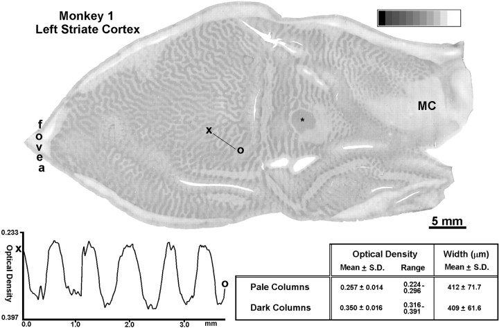
Fig. 2.
Monkey 1 (adult monocular enucleation). Single CO section of a flat-mount showing the ocular dominance columns in layer IVc 15 weeks after removal of the right eye. Measurements showed that the pale columns (right eye) and dark columns (left eye) in opercular cortex (left half of the tissue section) were virtually equal in width (see table). This was confirmed by a linear density profile, sampled across five sets of ocular dominance columns from X to O, which showed equal spacing in the peaks of optical density. For this plot the section was imaged at 2400 dpi to achieve a resolution of 10.58 μm/pixel. The mean optical density of the dark and light columns differed by 0.093. The complete montage of the ocular dominance columns in this animal has been published (Horton and Hocking 1996b, Monkey 2). *Blind spot representation of the right eye; MC, monocular crescent.

