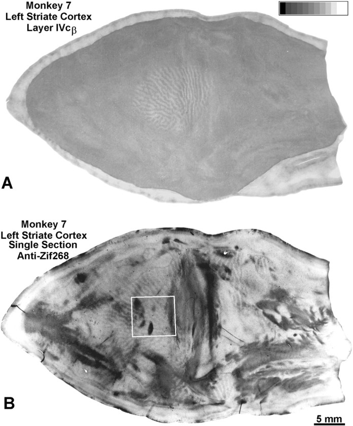
Fig. 9.

Monkey 7 (laser vs normal). A, Montage showing the scotoma induced by the laser lesion in Figure 8. Within the scotoma, the CO pattern resembles ocular dominance columns, proving that retinal damage sparing the ganglion cell layer still produces a CO pattern tantamount to enucleation. Measurements confirmed significant loss of CO activity, within both the lasered left eye’s ocular dominance columns (optical density, 0.351) and the intact right eye’s ocular dominance columns (optical density, 0.435), compared with CO activity outside the scotoma (optical density, 0.465). Outside the scotoma, CO staining was essentially homogeneous, because both retinas were normal, although there was some irregular fluctuation in density. This occurs as an artifact of montaging, because CO staining intensity varies slightly with section depth in layer IVcβ. B, Single Zif268 section showing ocular dominance columns in layer IVc. Outside the cortical scotoma, the dark Zif268 columns correspond to the intact left eye’s ocular dominance columns (Chaudhuri et al., 1995). Within the cortical scotoma, the dark Zif268 columns match the dark CO columns serving the freshly enucleated right eye. This is shown for theboxed region in Figure 10.
