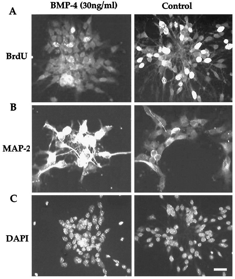
Fig. 1.
Representative examples of control and BMP-treated cultures. Immunostaining shows that BMPs promote neuronal differentiation and inhibit the number of cells that re-enter the cell cycle. A, There is a marked reduction in the number of cells incorporating BrdU after BMP-4 treatment relative to that in the control. B, More cells are positively stained for the neuronal marker MAP-2 after BMP-4 treatment than in untreated cultures. These positively stained cells have extensive dendritic trees typical of morphologically differentiated cortical neurons. C, DAPI nuclei staining shows comparable condensed nuclei and cluster size in control and BMP-4–treated cell cultures. Scale bar, 20 μm.

