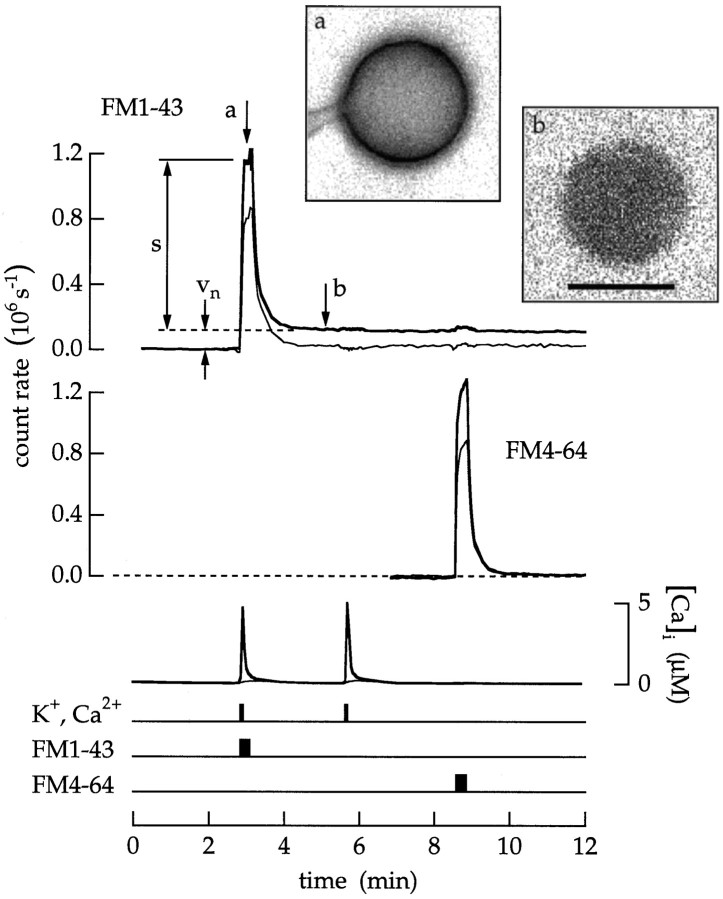
Fig. 10.
Endocytosis was induced by depolarization and Ca2+ influx. A bipolar cell was superfused with an extracellular saline containing 100 nmCa2+. Timing traces indicate an 18 sec application of 20 μm FM1–43, an 18 sec application of 20 μm FM4–64, and 6 sec applications of a high Ca2+ and K+ saline.s is the fluorescence intensity of the surface membrane.vn is the fluorescence of endocytosed vesicles that remain trapped in the cytoplasm. Transient endocytosis was measured as Tn = ρvn/s. After superfusion with the high Ca2+ and K+ saline,Tn was 0.08 MEq. Insets, Images of the terminal taken at the times marked a andb are shown. The gain in b is eight times the gain in a. Scale bar, 10 μm.

