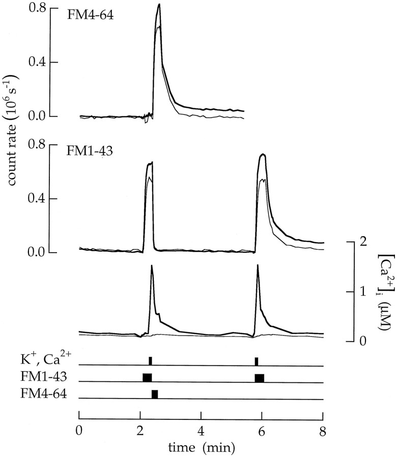
Fig. 13.
Endocytosis was delayed until after Ca2+ influx stopped. The timing of endocytosis was measured with 18 sec applications of 20 μm FM1–43 combined with 6 sec pulses of high Ca2+ and K+ saline. The first applications of FM1–43 and high Ca2+ and K+ saline ended together and were immediately followed by a pulse of 20 μm FM4–64 in a saline containing 100 nmCa2+. The FM4–64 quenched the fluorescence of FM1–43 in the surface membrane (see Fig. 3) but not in vesicles trapped in the cytoplasm. Tn measured (as shown in Fig. 10) with FM1–43 was 0.004 MEq and indicated that little endocytosis occurred during superfusion with the high Ca2+ and K+ saline.Tn measured with FM4–64 was 0.05 MEq and indicated that significant endocytosis occurred after return to the control saline. The second application of FM1–43 measured the total amount of endocytosis; Tn = 0.07 MEq.

