
Fig. 1.
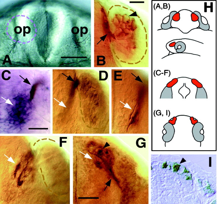
The pioneer neurons initiate the formation of the olfactory nerve and establish glomerular-like structures in the telencephalon. A, B, Frontal views with dorsal to the top. C–F, Dorsal views with anterior to the top. G, Ventral view with dorsal to the top. H, Diagrams labeled with letters corresponding topanels to show orientation. The second diagram is a side view, anterior to the left at a stage equivalent to the frontal view in H (A, B). The olfactory placodes are in red, and the eyes are ingray. I, A transverse section with anterior to the top. A, Frontal view of the head of a 24 h live zebrafish embryo. The olfactory placodes (op) are paired thickenings lying dorsoanteriorly. Thebroken line outlines the placode on theleft. B, Enlargement of a frontal view of a placode labeled with the zns-2 antibody at 24 h. Dendrites are absent on the apical surface (arrowhead), and the axons (arrow) exit basally and grow along the telencephalon. The broken line demarcates the edge of the placode.C, The pioneer axons (brown-blue,black arrow) meet the telencephalon in the region expressing the gene emx1 (purple,white arrow) in a 26 h embryo. D,E, A 30 h preparation showing two focal planes, labeled only with zns-2. The axons exit the placode (black arrows) and defasciculate in the telencephalon (white arrows). The broken line demarcates the posterior edge of the placode. F, At 38 h, the pioneer axons (white arrow) start to form condensations in the telencephalon. G, Axons labeled with zns-2 (black arrow) extend into the developing olfactory bulb in a 52 h embryo. Note the axonal condensations (white arrow) evident in the CNS. H, The developing olfactory placode has moved from a dorsal to a more anterior location in front of the developing eye (see A, B). As a result of this morphogenetic movement, the zns-2-labeled axons are more easily viewed from the ventral side (G,I) as they project anteriorly into the telencephalon. I, A 7.5 μm Epon section through the olfactory organ and bulb at the same developmental stage asG, with the axonal condensations indicated by thearrowhead. Scale bars: A, 40 μm;B, 25 μm; C–F, 35 μm;G, I, 40 μm.