
Fig. 2.
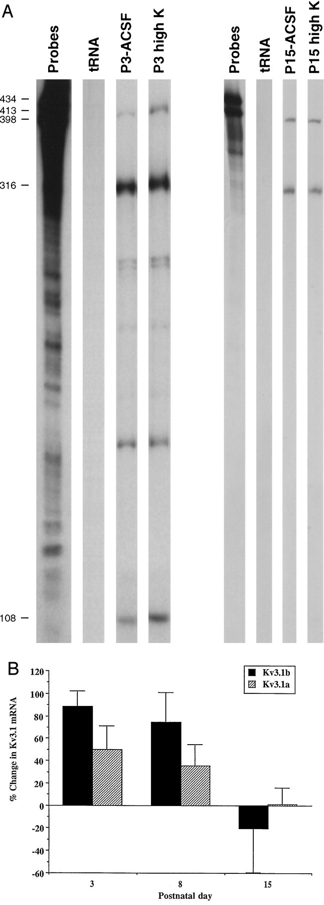
Modulation of Kv3.1 mRNA levels by depolarization during development. A, Effects of high-K treatment on the levels of the Kv3.1a and Kv3.1b mRNA, detected by an RNase protection assay, at P3 and P15. RNA was isolated from inferior colliculus slices incubated in ACSF and in high-K ACSF for 6 hr at room temperature. The bands corresponding to the Kv3.1b, Kv3.1a, and GAPDH mRNAs are 398, 108, and 316 nucleotides, respectively.B, Summary of the effects of depolarization onKv3.1a and Kv3.1b mRNA levels at P3, P8, and P15. Because there was variability in the specific activity of the GAPDH probe between experiments, the changes in the Kv3.1a and Kv3.1b mRNA levels were determined within each experiment. We calculated the average values of relative levels of Kv3.1a and Kv3.1b mRNA in the ACSF control sample and the changes in the Kv3.1a and Kv3.1b mRNA levels as follows:
All values are mean ± SEM. n values (the number of independent RNA preparations) at P3, P8, and P15 are 3, 4, and 2, respectively. The changes in Kv3.1b mRNA levels at P3 and P8 are significantly different from 0, p < 0.05 andp < 0.01, respectively, by a two-tailed Student’st test.