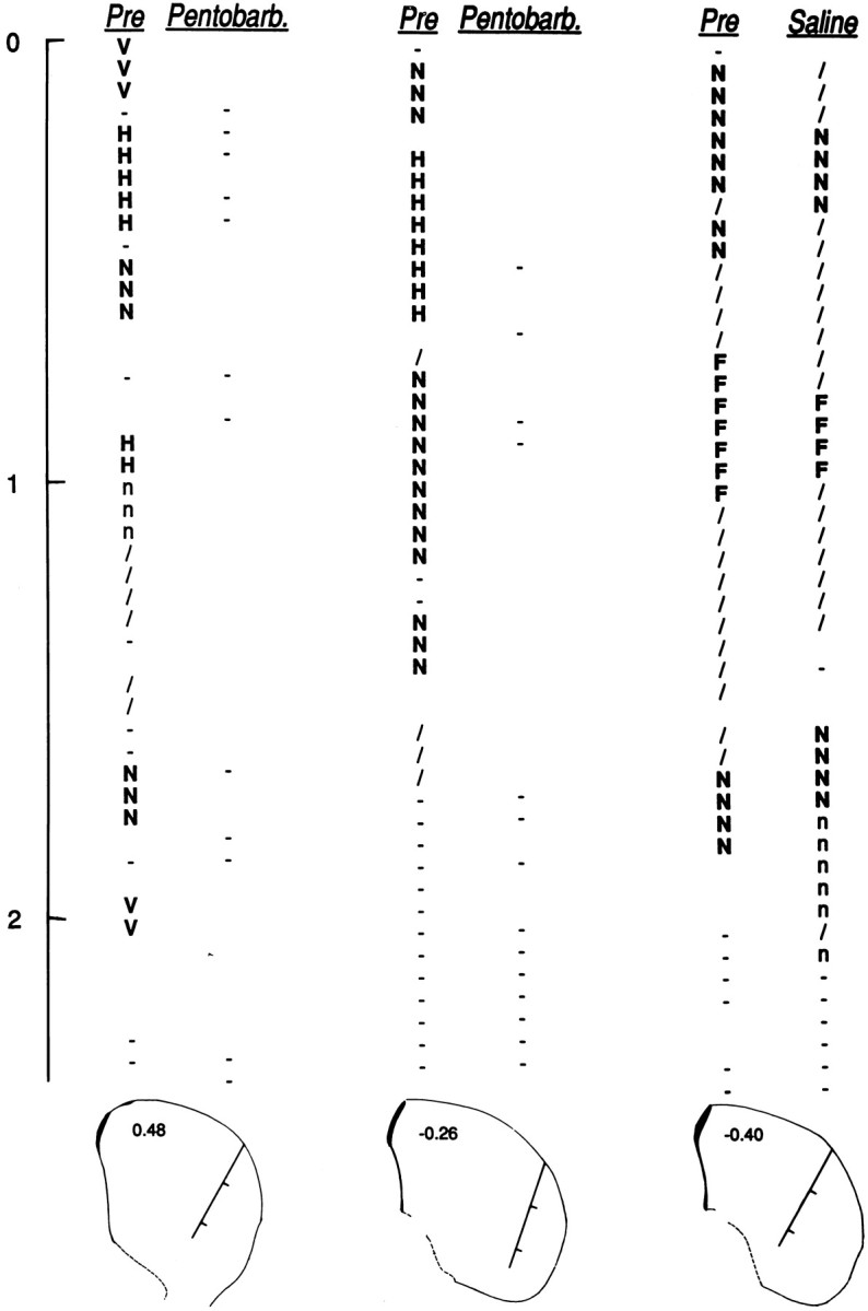
Fig. 2.

Three pairs of depth profiles through the lateral striatum. For each pair, penetrations were made along the same track before (Pre) and after injection of pentobarbital (Pentobarb.) or saline (Saline).Below each pair is a coronal diagram (Paxinos and Watson, 1996) nearest to the histologically verified location of the track. Tracks are marked in 1 mm divisions corresponding to thevertical scale at the far left (0 mm = point of entry into the striatum). Diagrams are labeled in millimeters anterior–posterior from bregma: 0.48 (left pair), −0.26, (middle pair), and −0.40 (right pair). A complete somatosensory exam was conducted every 50 μm. Uppercase letters indicate neurons responsive to somatosensory stimulation of individual body parts (F, forelimb; H, hindlimb;N, neck; V, vibrissae). Lowercase letters indicate neurons related only to active movement of a body part (n, neck); active-only firing was excluded from all analyses because it could not be compared between awake and anesthetized conditions. Slash, Neuron related to general movement; hyphen, neuron showing no evoked firing; blank space, no neuron recorded. Profiles were obtained from animals C39 (middle) and C44 (left and right).
