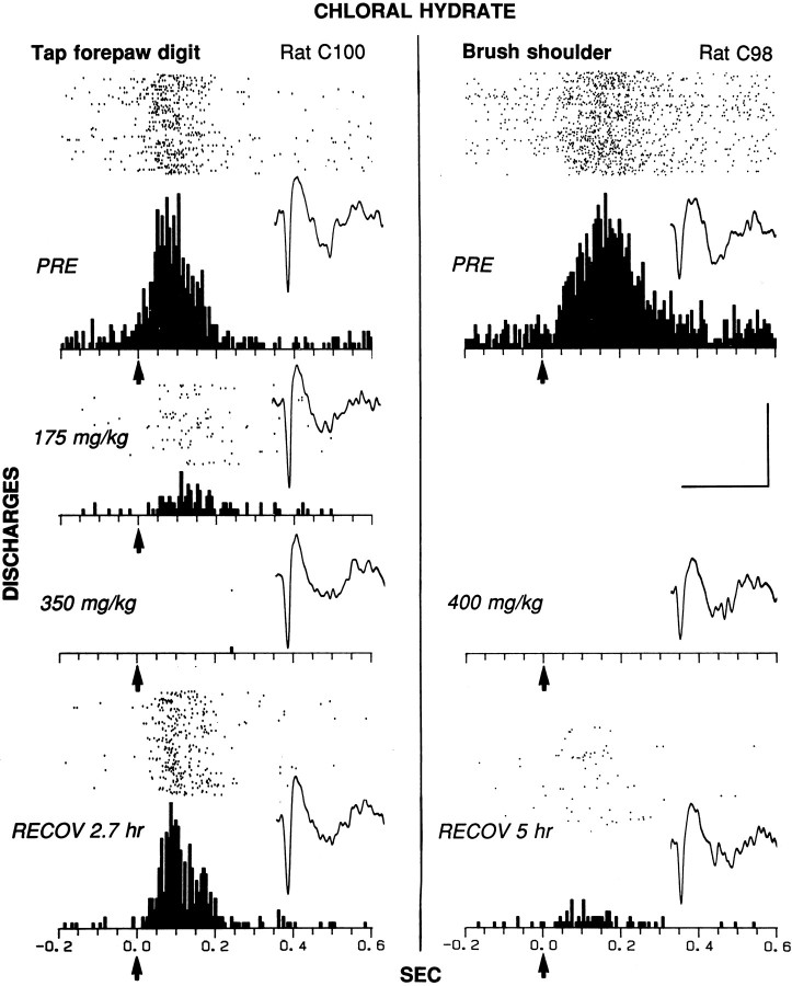
Fig. 5.
Elimination of cutaneous evoked discharges of two striatal neurons (left and right) in chronically implanted animals during chloral hydrate anesthesia. The raster, PSTH, and representative waveform of each neuron are shown for control (PRE) and for each dose and time period after injection. Left, The receptive field of the neuron included the whole contralateral forepaw up to the elbow in rat C100. The evoked response epoch was time 0 to 0.2 sec; the baseline epoch was −0.2 to 0 sec. The duration of the tap ranged between 0.1 and 0.2 sec; 75 sweeps were in each condition.Right, The neuron responded to soft brushing of the contralateral shoulder (brush tip, 5 mm × 10 mm; bristle length, 20 mm) in rat C98. The evoked response epoch was time 0 to 0.35 sec; the baseline epoch was −0.2 to 0 sec. The duration of the brush stroke was ∼0.3 sec; 90 sweeps were in each condition. Histological analysis verified that both neurons were located in the striatum, within 0.5 mm of its dorsolateral edge, as in the electrode track at thebottom left of Figure 2 (4.0 mm lateral to the midline; 0.5 mm anterior to bregma). Bin width = 6 msec. Eachtick on the x-axis = 50 msec.Arrows at time 0 indicate the approximate stimulus onset. Calibration (bars at right): for PSTHs, vertical bar, 20 discharges per bin; for waveforms, horizontal bar, 2.0 msec, and vertical bar, 0.15 mV (left) and 0.12 mV (right).

