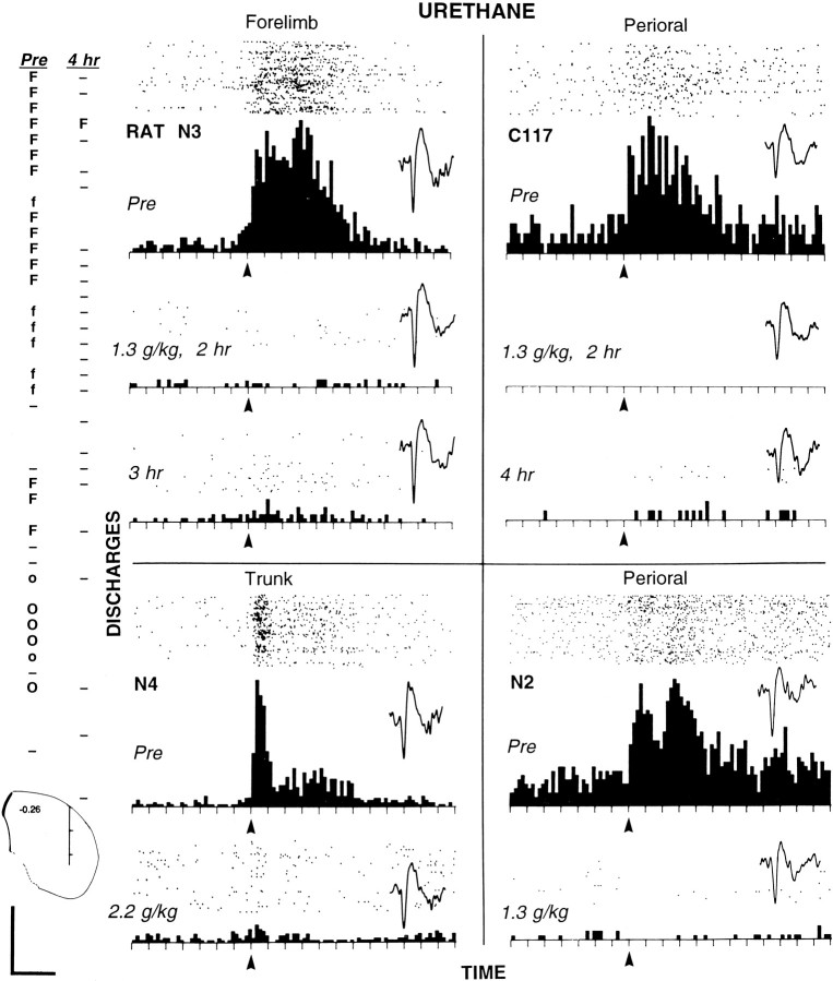
Fig. 6.
Elimination of somatosensory-evoked discharges of striatal neurons by urethane in four chronically implanted animals.Far left, Two electrode track profiles through the striatum (50 μm increments) obtained from rat N3 before (Pre) and 4 hr after injection of urethane (1.3 gm/kg).Pre profile (left) was obtained first; then the electrode was raised to the dorsalmost position to record the activity of the forelimb neuron (shown in top PSTH oftop left quadrant). Then urethane was injected, and the activity of the same forelimb neuron was recorded for the next 4 hr, after which the second profile (4 hr) was obtained. Below the profiles, a coronal diagram (Paxinos and Watson, 1996) shows their approximate location, −0.26 mm (anterior–posterior) and 4.0 mm (medial–lateral); hash marks are at each millimeter.Uppercase O, Neuron responsive to perioral stimulation;lowercase o, neuron correlated with licking;hyphen, neuron showing no discharges evoked by somatosensory stimulation. Details are described in the Figure 2legend. Four quadrants, Histograms (and rastersabove each) displaying the discharges of four neurons evoked by somatosensory stimulation in the four animals injected with urethane. Top panel in each quadrant, Awake state before injection. Lower panels ineach quadrant, Specific times after injection. Eachtick on the x-axis = 50 msec; bin width = 10 msec. Arrows indicate the approximate stimulus onset. For each of the four neurons, a representative waveform is shown from each time period for which a PSTH is displayed. Waveform calibration (bottom left): 0.2 mV; 1.0 sec. Top left quadrant (Figure legend continues),Forelimb neuron recorded at top location of profiles at far left in rat N3. (The neuron showed no evoked firing at 4 hr after injection but showed some evoked discharges in PSTH at 3 hr.) The evoked response epoch was 0 to 0.35 sec; the baseline epoch was −0.35 to −0.05 sec. Stimulus duration was ∼200 msec; 150 sweeps were in each condition. Top right quadrant, Neuron responsive to perioral stimulation recorded in rat C117. The evoked response epoch was 0 to 0.3 sec; the baseline epoch was −0.35 to −0.05 sec. Stimulus duration was ∼150 msec; 200 sweeps were in each condition. Lower left quadrant, Neuron responsive to cutaneous stimulation of the trunk in rat N4. The evoked response epoch was 0 to 0.3 sec; the baseline epoch was −0.35 to 0 sec. Stimulus duration was ∼100 msec; 200 sweeps were in each condition. Lower right quadrant, Neuron responsive to cutaneous stimulation of the perioral area in rat N2. The evoked response epoch was 0 to 0.25 sec; the baseline epoch was −0.35 to 0 sec. Stimulus duration was ∼150 msec; 100 sweeps were in each condition.

