
Fig. 1.
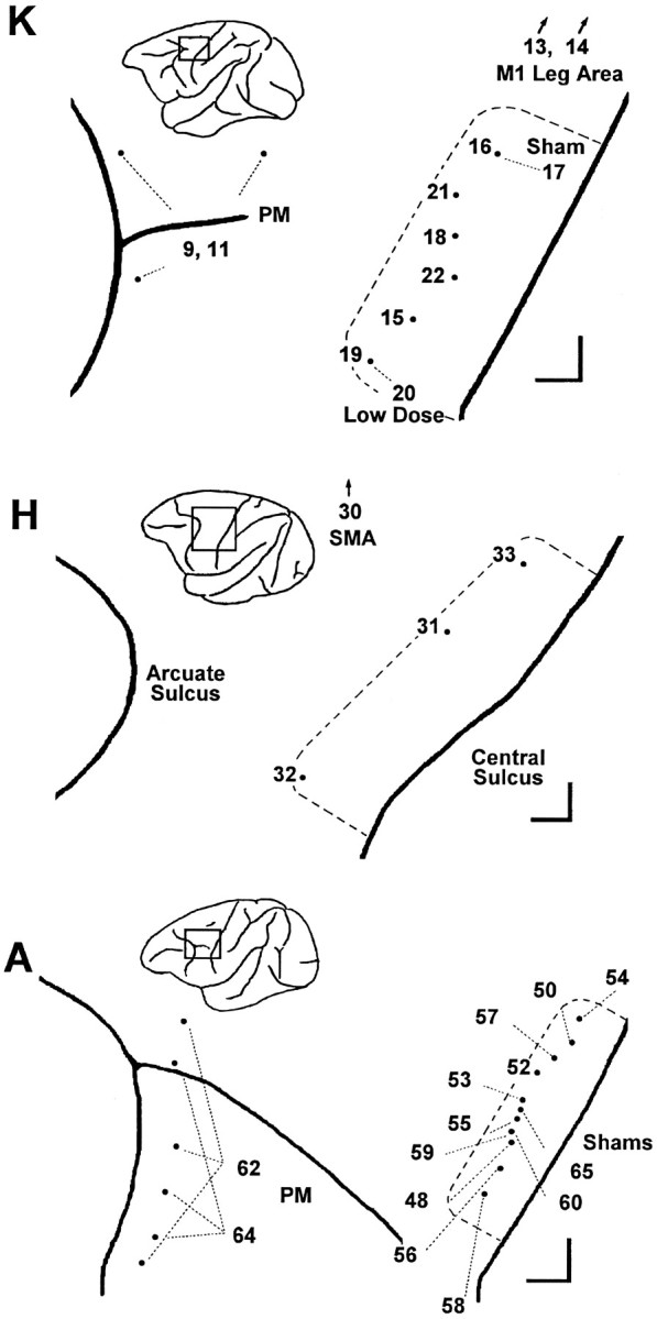
Location of injections in each session. For each monkey, K, H, and A, the sites of muscimol injections are shown aspoints on an enlarged view of the left hemisphere, with the central sulcus to the viewer’s right and the arcuate sulcus to the left. A rectanglein the inset of each monkey’s left hemisphere shows the region enlarged. Session numbers are shown either next to the point representing the injection site or else connected to the point with a fine dashed line. The extent of the M1 hand area in each monkey, as assessed with single-neuron recording and ICMS, is indicated by a heavy dashed line. Scale bars at thebottom right of each enlargement indicate 1 mm in the rostral and medial directions. In addition to muscimol injections in the M1 hand area, comparison control injections were made in the in the PM just posterior to the arcuate sulcus, in the M1 leg area, and in the SMA. M1 leg area sites and SMA sites were medial to the region shown enlarged (arrows). In PM, multiple sites were injected in the same session (see Table 1).