
Fig. 1.
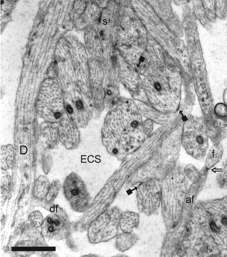
Representative CA1 neuropil from postnatal day 1 (R53) has large amounts of extracellular space (ECS). A dendrite (D) with well organized microtubules gives rise to a dendritic filopodium (df), which continues on adjacent serial sections. An asymmetric synapse (s) on a stubby dendritic protrusion is evident at the top of the figure. Also in evidence are nonsynaptic surface specializations (solid square arrows). The tip of an axonal filopodium (af), identified by tracing it back to its axonal origin, has a surface specialization or possibly a nascent synapse (open arrow) where it contacts the tip of a dendritic filopodium (f), also identified through series. Scale bar, 1 μm.