
Fig. 2.
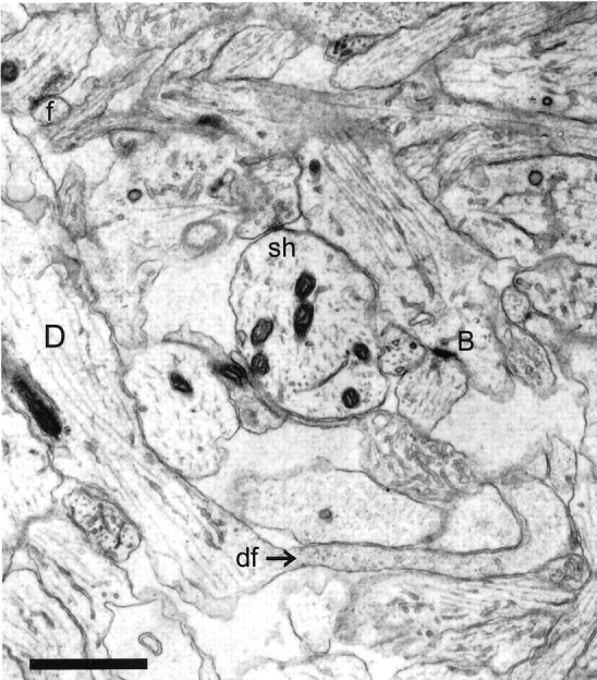
Representative CA1 neuropil from postnatal day 4 (R48a). A dendritic filopodium (df) emerges from the middle of a dendrite (D). The entire filopodium is reconstructed in Figure 5. A synapse on an apparently stubby profile is actually at the base (B) of a filopodium that could be traced on adjacent serial sections. A shaft synapse (sh) has synaptic vesicles in a docked position, suggesting the synapse is functional. The profile in the upper left corner (f) makes synaptic contact with an axon containing docked vesicles. This synapse occurs in the middle of a dendritic filopodium that could be followed for 70 sections to its tip, at which there was no synaptic contact. Scale bar, 1 μm.