
Fig. 3.
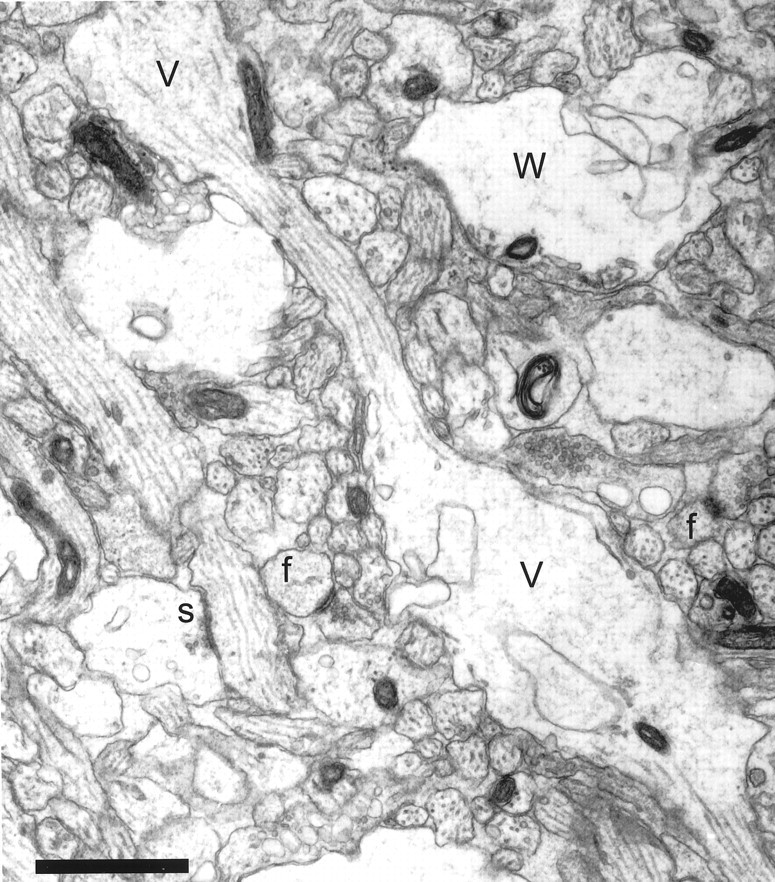
Neuropil from postnatal day 6 (R43b) showing varicosities (V) with interposed thin regions that give some dendrites a beaded appearance. Some varicosities had a watery cytoplasm (W) with organelles compressed to the periphery, suggesting that they might have undergone swelling. A synapse (s) located on a dendritic shaft has the appearance of a symmetric synapse with equally thin densities on both membranes. Asymmetric synaptic contacts on two dendritic filopodia profiles (f) were identified through serial sections. Note that these profiles might be mistaken for dendritic spines if they were viewed only on this section. Scale bar, 1 μm.